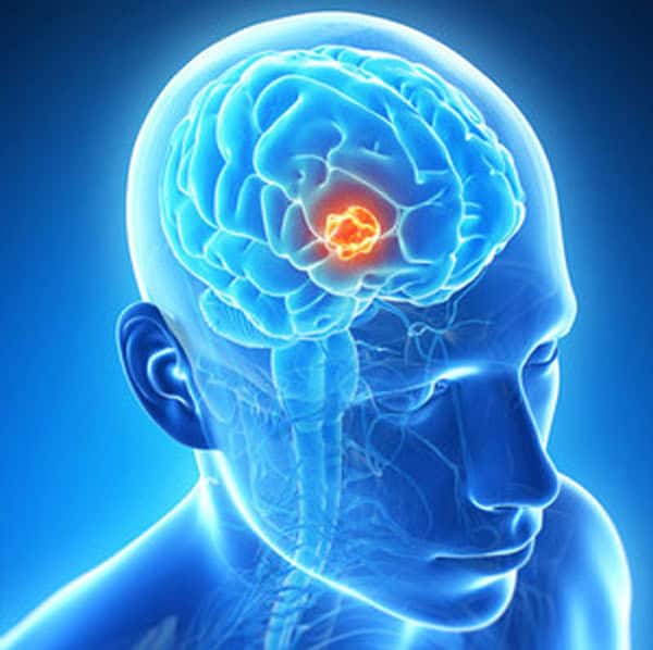
BRAIN TUMOR:
Primary brain tumors can be either malignant (contain cancer cells) or benign (do not contain cancer cells). A primary brain tumor is a tumor which begins in the brain. If a cancerous tumor which starts elsewhere in the body sends cells which end up growing in the brain, such tumors are then called secondary or metastatic brain tumors. This discussion is focused on primary brain tumors.
Brain tumors can occur at any age.

BRAIN TUMOR CAUSES:
Malignant glioma cells
Malignant glioma cells
Illustration showing acoustic neuroma
Acoustic neuroma (schwannoma)
Brain tumors that begin in the brain
Primary brain tumors originate in the brain itself or in tissues close to it, such as in the brain-covering membranes (meninges), cranial nerves, pituitary gland or pineal gland.
Primary brain tumors begin when normal cells acquire errors (mutations) in their DNA. These mutations allow cells to grow and divide at increased rates and to continue living when healthy cells would die. The result is a mass of abnormal cells, which forms a tumor.
Primary brain tumors are much less common than are secondary brain tumors, in which cancer begins elsewhere and spreads to the brain.
Many different types of primary brain tumors exist. Each gets its name from the type of cells involved. Examples include:
Gliomas: These tumors begin in the brain or spinal cord and include astrocytomas, ependymoma, glioblastomas, oligoastrocytomas and oligodendrogliomas.
Meningiomas: A meningioma is a tumor that arises from the membranes that surround your brain and spinal cord (meninges). Most meningiomas are noncancerous.
Acoustic neuromas (schwannomas): These are benign tumors that develop on the nerves that control balance and hearing leading from your inner ear to your brain.
Pituitary adenomas: These are mostly benign tumors that develop in the pituitary gland at the base of the brain.
These tumors can affect the pituitary hormones with effects throughout the body.
Medulloblastomas: These are the most common cancerous brain tumors in children. A medulloblastoma starts in the lower back part of the brain and tends to spread through the spinal fluid. These tumors are less common in adults, but they do occur.
PNETs: Primitive neuroectodermal tumors (PNETs) are rare, cancerous tumors that start in embryonic (fetal) cells in the brain. They can occur anywhere in the brain.
Germ cell tumors. Germ cell tumors may develop during childhood where the testicles or ovaries will form. But sometimes germ cell tumors move to other parts of the body, such as the brain.
Craniopharyngiomas: These rare, noncancerous tumors start near the brain’s pituitary gland, which secretes hormones that control many body functions. As the craniopharyngioma slowly grows, it can affect the pituitary gland and other structures near the brain.
Cancer that begins elsewhere and spreads to the brain
Secondary (metastatic) brain tumors are tumors that result from cancer that starts elsewhere in your body and then spreads (metastasizes) to your brain.
Secondary brain tumors most often occur in people who have a history of cancer. But in rare cases, a metastatic brain tumor may be the first sign of cancer that began elsewhere in your body.
Secondary brain tumors are far more common than are primary brain tumors.
Any cancer can spread to the brain, but the most common types include:
Breast cancer
Colon cancer
Kidney cancer
Lung cancer
Melanoma
Risk factors:
In most people with primary brain tumors, the cause of the tumor is not clear. But doctors have identified some factors that may increase your risk of a brain tumor. Risk factors include:
Your age. Your risk of a brain tumor increases as you age. Brain tumors are most common in older adults. However, a brain tumor can occur at any age. And certain types of brain tumors occur almost exclusively in children.
Exposure to radiation. People who have been exposed to a type of radiation called ionizing radiation have an increased risk of brain tumor. Examples of ionizing radiation include radiation therapy used to treat cancer and radiation exposure caused by atomic bombs.
More common forms of radiation, such as electromagnetic fields from power lines and radiofrequency radiation from cellphones and microwave ovens, have not been proved to be linked to brain tumors.
Family history of brain tumors. A small portion of brain tumors occur in people with a family history of brain tumors or a family history of genetic syndromes that increase the risk of brain tumors.

Symptoms:
The signs and symptoms of a brain tumor vary greatly and depend on the brain tumor’s size, location and rate of growth.
General signs and symptoms caused by brain tumors may include:
New onset or change in pattern of headaches
Headaches that gradually become more frequent and more severe
Unexplained nausea or vomiting
Vision problems, such as blurred vision, double vision or loss of peripheral vision
Gradual loss of sensation or movement in an arm or a leg
Difficulty with balance
Speech difficulties
Confusion in everyday matters
Personality or behavior changes
Seizures, especially in someone who doesn’t have a history of seizures
Hearing problems
Can cancer cells spread from one person to another?
Blood and cancer cells
Cancer spreading from one individual to another via blood is “very unlikely”
Can tumour cells get passed to another person through blood contact, for example from blood donations or used
needles, and can cancer be transmitted from the mother to an unborn child while in the womb?
When cancer in one part of the body spreads to another part of the body, the outlook for a patient is rarely positive.
Given how frequently this happens, it may come as a surprise to know that the spread of cancer from one person to another is actually incredibly rare.
“Generally, in well people who are not immune-suppressed, getting cancer from one individual to another via blood is very unlikely,”
“One study looked at about one-third of a million blood recipients, of which about 12,000 were at risk of being transfused blood from a donor with sub-clinical cancer and they found no increase in risk,”
This evidence fits with what we know about how the immune system responds to foreign matter. In the case of blood transfusion, blood type (such as A, B, AB and O) is carefully matched between the donor and recipient so the recipient’s immune system doesn’t see the red blood cells as foreign and destroy the red blood cells.
If there are cancer cells in that blood, there are other unique proteins on the surface of those cells that in the majority of cases, mark them out as foreign. The recipient’s immune system therefore identifies them and destroys them before they can settle in.
Blood banks also carefully screen donors to rule out anyone who’s had cancer, just in case.
But if the recipient’s immune system isn’t working well — for example, if they are immune-suppressed by illness or because they have had an organ transplant which requires immunosuppression of the recipient to prevent rejection of the donor organ — then they are less likely to be protected by this mechanism.
“When we do blood transfusion into immunocompromised people, we can irradiate the actual red cell units,” says Ng. This is done already to reduce the risk of the transfused white blood cells attacking the recipient’s body — something called graft versus host disease. This irradiation can also kill any sub-clinical cancer cells which may be circulating in the donor’s blood.
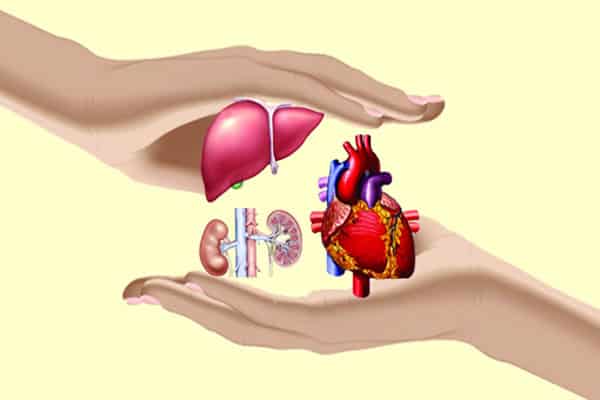
Organ donation and pregnancy:
In the case of solid organ transplants, such as liver or kidneys, there have been reports of cancer being unknowingly transmitted from the donor to the recipient. While donors and their organs are screened for cancer it can, on very rare occasions, slip through undetected. However the risk is incredibly low — around 0.015 per cent,
Finally, there is also evidence that cancer can be transmitted from a mother to her unborn child but again, this is very rare.
“In review back in 2003, there were only 14 reported cases in the literature where the mum had a type of cancer and the child also developed the same cancer,”. The cancers documented generally included aggressive types cancer and unfortunately likely to be during advanced stages of disease in the mother. Such cancers included
leukaemia, melanoma, solid organ cancers such as lung, and sarcomas.”
Transmission between mother and foetus can occur because of the unusual immunological relationship that exists between the two during pregnancy — one in which the foetal immune system is still relatively immature and may tolerate foreign cells. However, transmission of a maternal cancer to the foetus is very unlikely as this requires the cancer cells to be travelling in the mother’s circulation, and in addition, cross the placental barrier to the foetus. In most pregnancies, unless this placental barrier is breached such as with accidental trauma, foetal circulation remains completely separate from the mother’s blood supply.
Then there are the very rare, very unusual cases of person-to-person transmission, such as the surgeon who contracted cancer from a patient after accidentally cutting himself during surgery, and transmission of colon cancer through a needlestick injury.
In the case of the surgeon, it turned out that the cancer itself had performed a genetic miracle and incorporated some of the surgeon’s genes into itself. Dr Ng suggests that the explanation for these rare cases could also be related to immunologically similarities between the donor and recipient, or that the cancer cells somehow were able to evade immune detection.

Brain Tumor – Diagnosis:
Doctors use many tests to diagnose a brain tumor, find out the type of brain tumor, and rarely, find out if it has spread to another part of the body, called metastasis. Some tests may also determine which treatments may be the most effective. For most types of tumors, taking a sample of the tumor tissue, either by biopsy (see below) or by removing part or all of the tumor, is the only way to make a definitive diagnosis of a brain tumor. If this is not possible, the doctor may suggest other tests that will help make a diagnosis.
Imaging tests may be used to help determine whether the tumor is a primary brain tumor or if it is another type of cancer that has spread to the brain from elsewhere in the body. Your doctor may consider these factors when
Choosing a diagnostic test:
Age and medical condition
Type of tumor suspected
Signs and symptoms
Previous test results
Most brain tumors are not diagnosed until after symptoms appear. Often a brain tumor is initially diagnosed by an internist or a neurologist. An internist is a doctor who specializes in treating adults. A neurologist is a doctor who specializes in problems with the brain and central nervous system.
In addition to asking the patient for a detailed medical history and doing a physical examination, the doctor may recommend the tests described below to determine the presence, and perhaps the type or grade, of a brain tumor.
This list describes options for diagnosing a brain tumor, and not all tests listed will be used for every person. Based on the combined results of the different tests, the doctor will recommend treatment options.
Imaging tests:
The most effective and common tool for diagnosing a brain tumor is the use of a magnetic resonance imaging (MRI) scan, although computed tomography (CT or CAT) scans are also used. A positron emission tomography (PET) scan is used at first to find out more about a tumor while a patient is receiving treatment or if the tumor comes back after treatment.
Once an imaging scan shows that there is a tumor in the brain, the most common way to determine the type of brain tumor is to look at the results from a sample of tissue after a biopsy or surgery .
Each imaging test can provide specific information, but they must be combined with the results of the patient’s medical history, physical examination, and neurologic and other tests. The most common imaging tests used for diagnosing a brain tumor include:
MRI: An MRI uses magnetic fields, not x-rays, to produce detailed images of the body. MRI can also be used to measure the tumor’s size. A special dye called a contrast medium is given before the scan to create a clearer picture. This dye can be injected into a patient’s vein or given as a pill to swallow. MRIs create more detailed pictures than CT scans (see below) and are the preferred way to diagnose a brain tumor. The MRI may be of the brain, spinal cord, or both, depending on the type of tumor suspected and the likelihood that it will spread in the CNS. There are different types of MRI. The results of a neuro-examination, done by the internist or neurologist, helps determine which type of MRI to use.
Intravenous (IV): Gadolinium-enhanced MRI is typically used to help create a clearer picture of a brain tumor. This is when a patient first has a regular MRI, and afterwards is given a special type of contrast medium called gadolinium through an IV; a second MRI is then done to get another series of pictures using the dye.
A spinal:MRI may be used to diagnose a tumor on or near the spine.
A functional MRI (fMRI): Provides information about the location of specific areas of the brain that are responsible for muscle movement and speech. During the fMRI examination, the patient is asked to do certain tasks that cause changes in the brain and can be seen on the fMRI image. This test is used to help plan surgery, so the surgeon can avoid damaging the functional parts of the brain while removing the tumor.
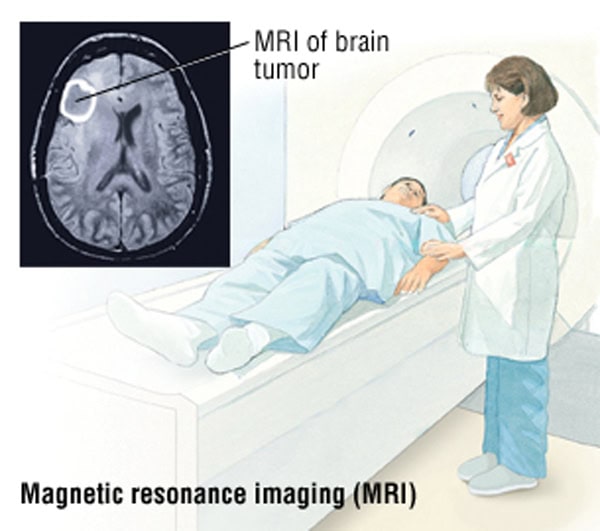
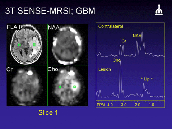
Magnetic resonance spectroscopy (MRS): Is a test using MRI that provides information on the chemical composition of the brain. It can help tell the difference between dead tissue caused by previous radiation treatments and new tumor cells in the brain.
CT scan:A CT scan creates a three-dimensional picture of the inside of the body with an x-ray machine. A computer then combines these images into a detailed, cross-sectional view that shows any abnormalities or tumors. A CT scan can help find bleeding and enlargement of the fluid-filled spaces in the brain, called ventricles. Changes to bone in the skull can also be seen on a CT scan, and it can be used to measure a tumor’s size. A CT scan may also be used if the patient cannot have an MRI, such as if the person has a pacemaker for his or her heart. Sometimes, a contrast medium is given before the scan to provide better detail on the image. This dye can be injected into a patient’s vein or given as a pill to swallow.
PET scan: A PET scan is a way to create pictures of organs and tissues inside the body. A small amount of a radioactive sugar substance is injected into the patient’s body. This sugar substance is taken up by cells that use the most energy. Because cancer tends to use energy actively, it absorbs more of the radioactive substance. A scanner then detects this substance to produce images of the inside of the body.
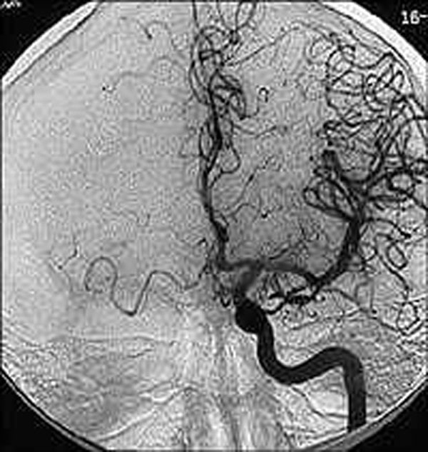
Cerebral arteriogram, also called a cerebral angiogram. A cerebral arteriogram is an x-ray, or series of x-rays, of the head that shows the arteries in the brain. X-rays are taken after a contrast medium is injected into the main arteries of the patient’s head.
Lumbar puncture or spinal tap: A lumbar puncture is a procedure in which a doctor uses a needle to take a sample of cerebrospinal fluid (CSF) to look for tumor cells, blood, or tumor markers. Tumor markers or biomarkers are substances found in higher than normal amounts in the blood, urine, spinal fluid, plasma or other bodily fluids of people with certain types of cancer. Typically a local anesthetic is given to numb the patient’s lower back before the procedure.
Myelogram: Because some specific types of brain tumors can spread to the spinal fluid, other parts of the brain, or the spinal cord, the doctor may recommend a myelogram to look for areas where the tumor may have spread. A myelogram uses a dye injected into the CSF that surrounds the spinal cord. The dye shows up on an x-ray and can outline the spinal cord to help the doctor look for a tumor. This is rarely done; a lumbar puncture (see above) is more common.
Tissue sampling/biopsy/surgical removal of a tumor:
As explained above, imaging tests are useful, but a sample of the tumor’s tissue is usually needed for the final diagnosis. A biopsy is the removal of a small amount of tissue for examination under a microscope and is the only definitive way a brain tumor can be diagnosed. A pathologist then analyzes the sample(s). A pathologist is a doctor who specializes in interpreting laboratory tests and evaluating cells, tissues, and organs to diagnose disease. A biopsy can be done as part of surgery to remove the entire tumor or as a separate procedure if surgical removal of the tumor is not possible because of its location or a patient’s health.
Molecular testing of the tumor:
Your doctor may recommend running laboratory tests on a tumor sample to identify specific genes, proteins, and other factors, such as tumor markers, unique to the tumor. Some biomarkers may help doctors determine a patient’s prognosis (see Grades and Prognostic Factors). Researchers are examining biomarkers to find ways to diagnose a brain tumor before symptoms begin. Ultimately, results of these tests may help decide whether your treatment options include a type of treatment called targeted therapy .
Neurological, vision, and hearing tests:
These tests help determine if a tumor is affecting how the brain functions. An eye examination can detect changes to the optic nerve, as well as changes to a person’s field of vision.
Neurocognitive assessment
This consists of a detailed assessment of all major functions of the brain, such as storage and retrieval of memory, expressive and receptive language abilities, calculation, dexterity, and the overall well-being of the patient. These tests are done by a licensed clinical neuropsychologist, who will write a formal report to be used for comparison with future assessments or to identify specific problems that can be helped through treatment.
Electroencephalography (EEG):
An EEG is a noninvasive test in which electrodes are attached to the outside of a person’s head to measure electrical activity of the brain. It is used to monitor for possible seizures .
Evoked potentials:
Evoked potentials involve the use of electrodes to measure the electrical activity of nerves and can often detect acoustic schwannoma, a noncancerous brain tumor. This test can be used as a guide when removing a tumor that is growing around important nerves.
Test results:
After diagnostic tests are done, your doctor will review all of the results with you. If the diagnosis is a tumor, additional tests will be done to learn more about the tumor. The results help the doctor describe the tumor and plan treatment.
People with brain tumours have several treatment options as follows: surgery, radiotherapy, chemotherapy and other drug therapies.
Treatment for brain tumours is based on many factors such as:
Patient age, overall health and medical history
Type, location and size of the tumour
How likely the tumour is to spread or recur, or how fast the tumour is growing
Patient tolerance for specific medications, procedures or therapies
Decisions about the best course of treatment will be made together with the patient and their medical team—there is no ‘one-size-fits-all’ treatment option for brain cancer.
The medical team, also called a multidisciplinary team, can include neuro-oncologists, medical oncologists, radiation oncologists, surgeons, nurses, social workers, rehabilitation therapists, neuropsychologists, and other specialists.
In Australia, some people may be offered the option of participation in a clinical trial to test new ways of treating brain cancer. A clinical trial is a research study to test a new treatment to evaluate whether it is safe, effective, and possibly better than the standard treatment.
Standard treatments:
Surgery:
Surgery is usually the first treatment used for a brain tumour.
The aim of surgery is to remove as much of the tumour as possible, while minimising damage to surrounding healthy brain. Sometimes it is not safe or possible to remove all visible tumour tissue because it is too close to important areas of healthy brain.
Other aims of surgery are:To provide a large specimen for examination by the pathologist, to confirm the diagnosis and help guide treatment to relieve the pressure on the brain. This can improve symptoms and may reduce the amount of drugs the person needs to control symptoms to reduce the amount of remaining tumour to be treated with radiotherapy and chemotherapy. These treatments may be better tolerated and have less side effects if there is less tumour left to treat.
If the brain tumour is located near a part of the brain that controls speech, or movement or some other vital function, it is common to perform the operation when the patient is awake for a short part of the surgery. The patient is woken once the surface of the brain is exposed and special electrical stimulation techniques are used to locate the specific part of the brain that controls speech, movement, or vision. This avoids causing damage while removing the tumour.
Sometimes a tumour cannot be removed because it would be too dangerous. This is called an irresectable or unresectable tumour. In this case, the medical team will discuss other treatment options with the patient to ease symptoms.
Radiotherapy:
X-rays and other forms of radiation can destroy tumour cells or delay tumour growth. Radiotherapy is recommended for all people with high-grade tumours if they are well enough to have this treatment, because it can prolong their survival.
Radiotherapy starts as soon as possible after the diagnosis of high-grade tumour; usually two-to-six weeks after surgery, when the surgical wound has healed. Treatment generally takes place Monday to Friday for about six weeks.
Radiotherapy may be given when the tumour is growing or becoming more invasive, or when surgery is unsuitable.
Chemotherapy:
Chemotherapy is the use of drugs to kill cancer cells, usually by stopping the cancer cells’ ability to grow and divide.
The goal of chemotherapy can be to destroy cancer cells remaining after surgery, slow a tumour’s growth, or reduce symptoms. Chemotherapy is typically given after surgery and possibly along with radiotherapy. Chemotherapy drugs may be delivered intravenously or directly into the cerebrospinal fluid (CSF), via injection, or orally.
Chemotherapy is recommended for all patients with glioblastoma multiforme (GBM) who are well enough for the treatment. Chemotherapy normally begins at the same time as radiotherapy and normally continues for six months after radiotherapy. For people with lower grade tumours, such as grade III astrocytoma, if chemotherapy is used, it is given after radiotherapy.
In some cases, implants containing a chemotherapy drug are inserted during surgery into the cavity left after the visible tumour is removed.

Other treatments:
Radiosurgery:
Traditional forms of radiotherapy expose both healthy and tumour tissue to high doses of radiation to reduce tumour growth. Newer methods of radiotherapy – called radiosurgery – are highly precise, exposing only tumour tissue and minimal surrounding tissue to the radiation. Through precise targeting of the tumour higher doses of radiation can be used, reducing the number of doses needed and the risks and side effects associated with the treatment.
Cyberknife:
The CyberKnife, delivers multiple beams of x-rays using a robotic arm. It is image-guided so can adjust to the natural movements of the organs and work anywhere in the body. It is used for the treatment of certain lung, brain, spine, liver and prostate cancers which otherwise may be inoperable, or where other treatment options may compromise other vital organs. CyberKnife treatments are delivered in one session or can be staged over several days. Typically brain cancer treatments are completed within five days. For most patients the CyberKnife treatment is a completely pain-free experience.
Gamma Knife:
The Gamma Knife delivers gamma rays to a highly-defined target within the brain. The Gamma Knife utilises a lightweight frame to hold the head in place and provide a reference point for targeted radiosurgery. Imaging is performed prior to radiation treatment to ensure that the Gamma Knife’s beams are focused on the tumour site. Working together, neurosurgeons and radiation oncologists identify the target and develop a plan to deliver an extremely accurate dose of radiation while reducing exposure to sensitive healthy tissue. Treatment sessions can last from a few minutes to an hour.
Gamma Knife surgery is performed at Macquarie University Hospital in Sydney, New South Wales. The first Gamma Knife technology to be made available in a public hospital in Australia is at the Princess Alexandria Hospital in Queensland, with the first patients expected to be treated from September 2015.
Proton Beam therapy:
Proton beam therapy is an advanced form of radiotherapy which targets tumours with great precision and where the radiation dose can be significantly and safely increased to help eradicate the cancer. Protons are positively charged particles found in the nucleus of every atom. Protons are made available in this therapy by stripping away electrons from hydrogen atoms. As protons move through the body they slow down, causing greater damage to surrounding cells. Due to this unique property, Proton Therapy causes minimal damage to healthy tissue as it enters the body and almost no damage to tissue as it exits the body, unlike traditional radiation therapy. As a result, the radiation oncologist can increase the dose to the tumour while reducing the dose to surrounding normal tissues. Proton therapy is used in the treatment of certain solid cancers in children, tumours of the eye and base of skull, and is becoming the treatment of choice for cancers of the head and neck, brain and spine, prostate, lung, gastrointestinal tract and breast.
Other drug therapies:
Other drugs used to treat people with brain tumours include:
Pain medication to help manage the pain from headaches, a common symptom of a brain tumour. Often, drugs call corticosteroids are used to control pain without the need for prescription pain medications.
Anti-seizure medication to help control seizures. There are several types of drugs available.
Corticosteroids are also used to decrease the amount of swelling in the brain.
How to Prevent Brain Cancer:
Brain cancer is an attack of small tumors on the brain or close to it. There are many types of brain tumors that may be benign or malignant. In most cases, doctors do not know the cause of brain cancer, though they do know that there are certain risk factors that make you more susceptible to developing the disease.Doctors do not yet understand the major causes of the primary tumors. Although you may not definitively be able to control getting brain cancer, by understanding your risk factors, the disease, and taking proactive steps such as regular check ups and living a healthy lifestyle, you may decrease your risk of developing tumors or cancer of the brain or surrounding area.
Preventing the Development of Brain Cancer
Image titled Prevent Brain Cancer
Be ware of risk:
Doctors do not know what causes brain in most cases, but there are certain factors that can increase your risk.
Knowing these factors can help you identify your risk and potential symptoms, and get regular checkups.
The main risk factors for brain cancer include age, exposure to radiation, a family history of brain tumors, and currently having cancer that could metastasize, or spread, to your brain from another area of your body.
Image titled Prevent Brain Cancer
Recognize your risk increases with age.
Any person, from children to the elderly, can develop brain cancer. However, your risk for the disease increases the older you get. Recognizing this and being aware of your body may help you to seek a medical opinion if you notice any symptoms of brain cancer.
Some brain tumors and cancer, such as brainstem gliomas and astrocytomas, are almost exclusively present in children.
Image titled Prevent Brain Cancer
Ask about your family’s medical history:
Keep a detailed record of your family’s medical history, including cases of cancer and tumors. If you have a family history of brain tumors or certain genetic syndromes that increase risk for brain cancer, you are at a higher risk of developing cancer of the brain or surrounding areas. Understanding your family medical history of brain cancer can identify potential symptoms and treatment options.
It’s always wise to keep a personal record of your family’s medical history and to have one at your doctor’s office.
Only 5-10% of all cancers are hereditary and brain cancer comes from a genetic mutation passed down from grandparent to parent.
People and family members with Li-Fraumeni syndrome, neurofibromatosis, tuberous sclerosis and Turcot syndrome may be more susceptible to brain cancer.

Image titled Prevent Brain Cancer:
Limit exposure to radiation.
Different types of radiation can increase your risk of developing brain cancer. Limiting your exposure to radiation may help you prevent developing the disease.
Ionizing radiation, which is present in some radiation therapies for cancer or atomic bombs, increases your risk of brain cancer. You may not be able to limit your exposure to ionizing radiation if you are undergoing treatment for another cancer. The likelihood of being exposed through an atomic bomb or nuclear meltdown is low.
Ultraviolet radiation, which the sun emits, can also increase your risk for brain cancer. Wearing sunscreen and limiting sun exposure may decrease your risk.
Image titled Prevent Brain Cancer:
Understand what kinds of radiation do not cause brain cancer:
People are often exposed to more common forms of radiation including electromagnetic fields or radiofrequency radiation. Although some people believe that these types of radiation cause brain cancer, there is no evidence linking them to brain tumors.
Studies have not linked radiation from power lines, cellphones and microwaves to brain cancer.
Stay abreast of research on radiation exposure, which may help identify your risk factors.

Image titled Prevent Brain Cancer:
Change your eating and nutritional habits:
There is some evidence that nutritional habits during fetal development, during childhood and into adulthood may decrease your risk of developing brain cancer. Eating plenty of fruits and vegetables and lowering cholesterol may help you prevent brain cancer.
If your mother ate fruits and vegetables during her pregnancy and/ or gave them to you as a part of your diet during childhood, you may be at a lower risk of developing brain cancer.
Continuing to eat a diet rich in different fruits and vegetables may keep your risk for brain cancer lower.
Lowering your cholesterol and limiting how much fatty food you eat may minimize your risk for brain cancer.

Image titled Prevent Brain Cancer:
Exercise regularly:
Aim to exercise most days of the week. Doing cardiovascular exercise can help you stay healthy and may minimize your risk of developing brain cancer.
Aim to walk 10,000 steps a day, which translates to walking about 5 miles (or 8km) per day.Wearing a pedometer can help you make sure you’re taking enough steps per day.
You can do any type of cardio training to maintain your health. Beyond walking, consider running, swimming, rowing, or biking.


